VCBM 2016 paper ‘Sline: Seamless Line Illustration for Interactive Biomedical Visualization’ accepted!

Our paper ‘ Sline: Seamless Line Illustration for Interactive Biomedical Visualization’ was accepted for presentation at VCBM 2016, the 6th Eurographics Workshop on Visual Computing for Biology and Medicine. I’ve attended all VCBM editions since 2012, and am happy I can attend this one as well in Bergen, Norway. Which is extra convenient, since it’s my new hometown! I accepted a position as a researcher in the amazing visualization group at the University of Bergen and just started this week ^^

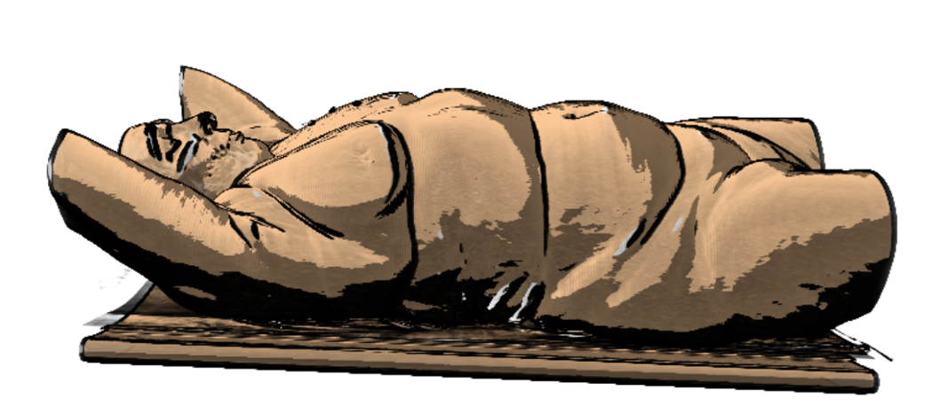
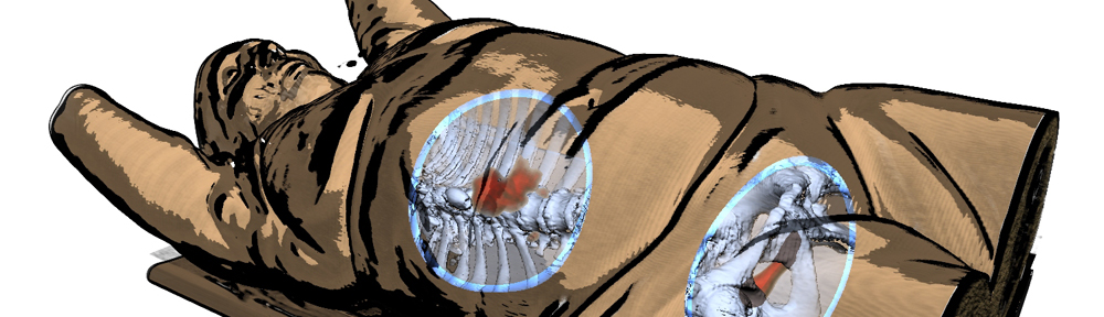
Back to Sline though, it’s a cool technique where you can pick an illustrative rendering style per structure using a single parameter slider. Behold:

Our Virtual Surgical Pelvis illustrated using Sline
Our Virtual Surgical Pelvis illustrated using Sline. So the organs and bones are hatched to draw attention here, while the nerves and vessels are de-emphasized by rendering only the silhouettes.

authors:  Nils Lichtenberg, Noeska Smit, Christian Hansen, and Kai Lawonn

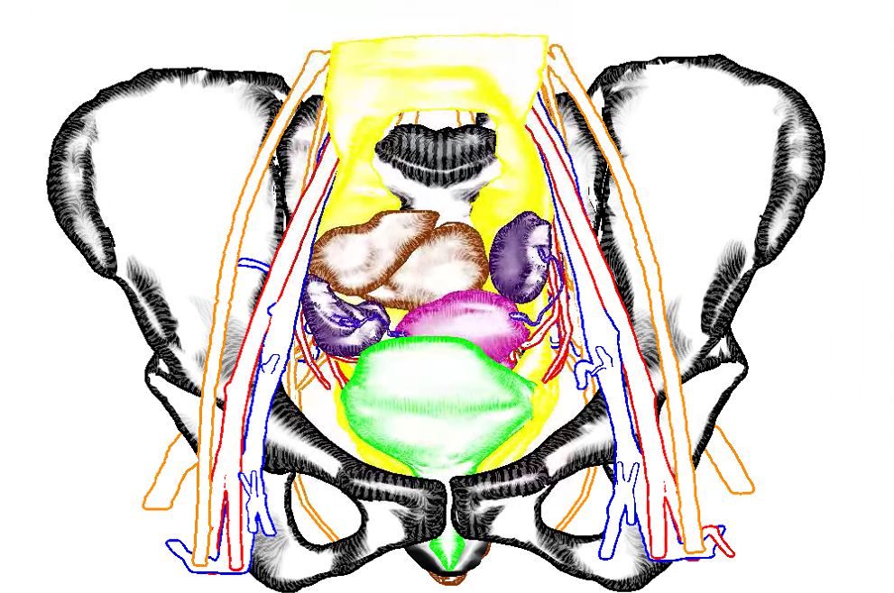
abstract: In medical visualization of surface information, problems often arise when visualizing several overlapping structures simultaneously. There is a trade-off between visualizing multiple structures in a detailed way and limiting visual clutter, in order to allow users to focus on the main structures. Illustrative visualization techniques can help alleviate these problems by defining a level of abstraction per structure. However, clinical uptake of these advanced visualization techniques so far has been limited due to the complex parameter settings required.

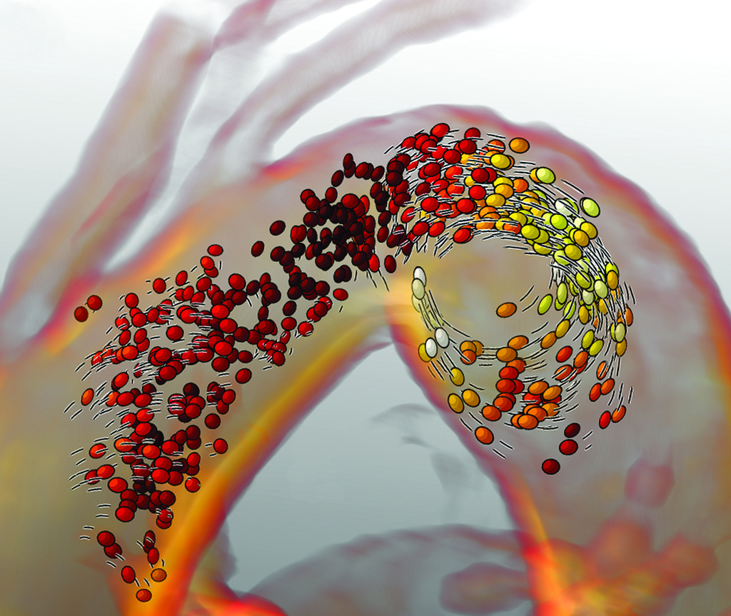
To bring advanced medical visualization closer to clinical application, we propose a novel illustrative technique that offers a seamless transition between various levels of abstraction and detail. Using a single comprehensive parameter, users are able to quickly define a visual representation per structure that fits the visualization requirements for focus and context structures. This technique can be applied to any biomedical context in which multiple surfaces are routinely visualized, such as neurosurgery, radiotherapy planning or drug design. Additionally, we introduce a novel hatching technique, that runs in real-time and does not require texture coordinates. An informal evaluation with experts from different biomedical domains reveals that our technique allows users to design focus-and-context visualizations in a fast and intuitive manner.

Comic Book-Inspired Illustrative Medical Visualization

Medical visualization isn’t always about rendering data in the most realistic way possible. In fact, quite often it isn’t. Illustrative rendering techniques have been developed that present medical data to the viewer in a style that is more abstract, often emphasizing important features, while taking emphasis away from the less relevant. Illustrative rendering techniques can even make data originating from medical imaging scanners such as CT, look hand-drawn. Check out for instance the works by Tobias Isenberg, Stefan Bruckner, Ivan Viola and Kai Lawonn to name a few. As you may recall, last year’s VCBM paper I had the pleasure of co-authoring also involved illustrative rendering:

Illustrative rendering of a CT scan

So what you see here kind of looks like a sketch hinting at the shape of the human body on a table right (well with some limbs missing ;))? Actually it’s a rendering of a CT scan combining toon shading and feature lines without any artists involved. Here’s another example by Roy van Pelt:

Roy van Pelt's blood flow visualization
Roy van Pelt’s blood flow visualization

He visualized blood flow using particles that get elongated as they move faster in combination with speed lines indicating the direction and speed of the particles. This style is reminiscent of something you could see in comic books or cartoons. In fact, I think there is a lot we can learn from comic book artists that we can apply to medical visualization.

For this reason, I asked Gerrit Rijken, AKA Iosua, a freelance illustrator as well as comic book aficionado, to write about the basics of inking, and more specifically the do’s and don’t’s and why’s of it all. In his elaborate post, which you can find here, he talks us through many things that are of interest in illustrative rendering as well. He describes how inking techniques are applied to create textures, the importance of line weights, spotting blacks, screen tones, feathering and cross-hatching. He provides explanations of these techniques combined with examples illustrating the concepts.

I see many parallels to visualizing techniques. For instance, focus-and-context techniques and depth cues are relevant for both comic books and medical visualizations. There are also some interesting rules on line thickness that I had not considered before. Furthermore I see techniques that have already been adopted in medical illustrative rendering, such as stippling and hatching, while I did not see researchers using feathering yet (correct me if I’m wrong ^^). I hope you find this as interesting as I did and if you have further questions, do not hesitate to contact him!

VCBM paper ‘Illustrative Multi-volume Rendering for PET/CT Scans’ accepted!

Our full paper on Illustrative Multi-volume Rendering for PET/CT Scans[1] was accepted for presentation at the always awesome VCBM workshop. I will be presenting it in Chester (UK) in September. For now, a teaser image of our technique:

lasmprvi_vcbm2015
Illustrative Multi-volume Rendering for PET/CT Scans teaser image