MedViz image contest 2016 winner!

noeska smit
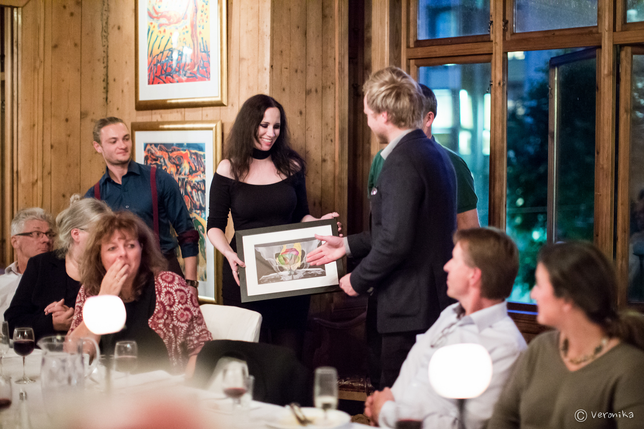
I know, it’s been a while since I last posted, and it’s also been a while since this happened, but… I was one of the winners of the MedViz image contest 2016 which was given out during the joint MedViz conference and Eurographics Workshop on Visual Computing in Biology and Medicine (aka VCBM), right here in Bergen.

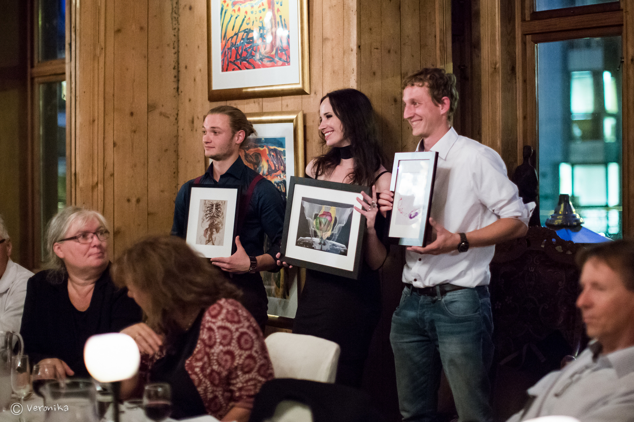
There were three winners in this image contest, and besides our pelvis, also Sergej Stoppel and Niels de Hoon received a prize, with their works entitled ‘Arteries in focus’ and ‘Turbulent flow in an aorta’, respectively:

medviz image contest 2016 winners
MedViz image contest 2016 winners

I sent in the following submission:

PelVis

PelVis
Authors
Noeska Smit, Kai Lawonn, Annelot Kraima, Marco de Ruiter, Hessam Sokooti, Stefan Bruckner, Elmar Eisemann, Anna Vilanova
Short Description
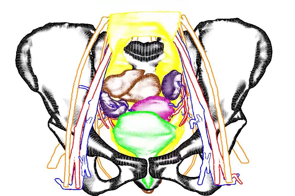
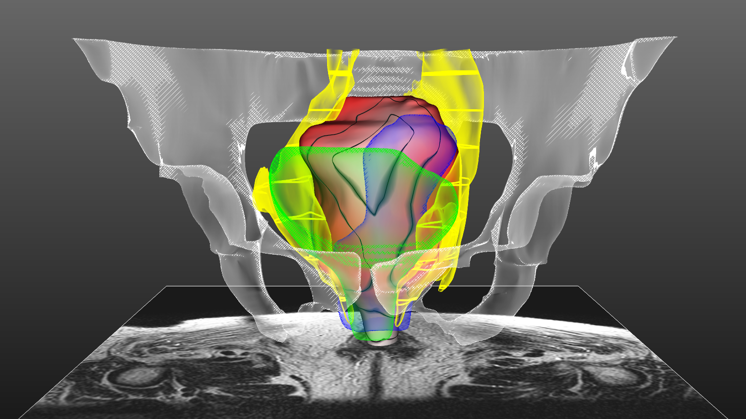
This image depicts PelVis [1], an interactive application for surgical planning for the Total Mesorectal Excision (TME) procedure. During this surgical procedure, undesired side-effects occur in up to 80% of the cases due to damage to the autonomic nerves. These nerves are damaged easily, since they are not visible in pre-operative MRI or even during the surgery. In order to visualize these nerves, we built an atlas model, the Virtual Surgical Pelvis (VSP) [1], that reveals zones in which the autonomic nerves reside based on cryosection and immunohistochemical studies. In the PelVis application, we register this atlas to patient-specific clinical MRI data and thus are able to make patient-specific virtual models of the individual patient, and to reveal the autonomic nerve zones pre-operatively, as displayed here in yellow. We highlight the distance of the mesorectal wall to these nerve zones using a colormap (red to white) combined with isolines. Furthermore, other surgically relevant anatomy is shown for spatial context, without occluding the view on the mesorectum, and the linked atlas-enriched MRI data can be explored interactively [3].
[1]: Smit, N., Lawonn, K., Kraima, A., DeRuiter, M., Sokooti, H., Bruckner, S., … & Vilanova, A. PelVis: Atlas-based Surgical Planning for Oncological Pelvic Surgery. (2017) IEEE Transactions on Visualization & Computer Graphics, (1), 1-1. Accepted, to appear.
[2]: Kraima, A., Smit, N. N., Jansma, D., West, N. P., Quirke, P., Rutten, H. J., … & DeRuiter, M. C. (2014). 62. The virtual surgical pelvis: A highly-detailed 3D pelvic model for anatomical education and surgical simulation. European Journal of Surgical Oncology, 40(11), S32.

That’s right, we now have an award-winning pelvis :). Pictures in this post were made by the multi-talented Veronika Šoltészová.

VCBM 2016 paper ‘Sline: Seamless Line Illustration for Interactive Biomedical Visualization’ accepted!

Our paper ‘ Sline: Seamless Line Illustration for Interactive Biomedical Visualization’ was accepted for presentation at VCBM 2016, the 6th Eurographics Workshop on Visual Computing for Biology and Medicine. I’ve attended all VCBM editions since 2012, and am happy I can attend this one as well in Bergen, Norway. Which is extra convenient, since it’s my new hometown! I accepted a position as a researcher in the amazing visualization group at the University of Bergen and just started this week ^^

Back to Sline though, it’s a cool technique where you can pick an illustrative rendering style per structure using a single parameter slider. Behold:

Our Virtual Surgical Pelvis illustrated using Sline
Our Virtual Surgical Pelvis illustrated using Sline. So the organs and bones are hatched to draw attention here, while the nerves and vessels are de-emphasized by rendering only the silhouettes.

authors:  Nils Lichtenberg, Noeska Smit, Christian Hansen, and Kai Lawonn

abstract: In medical visualization of surface information, problems often arise when visualizing several overlapping structures simultaneously. There is a trade-off between visualizing multiple structures in a detailed way and limiting visual clutter, in order to allow users to focus on the main structures. Illustrative visualization techniques can help alleviate these problems by defining a level of abstraction per structure. However, clinical uptake of these advanced visualization techniques so far has been limited due to the complex parameter settings required.

To bring advanced medical visualization closer to clinical application, we propose a novel illustrative technique that offers a seamless transition between various levels of abstraction and detail. Using a single comprehensive parameter, users are able to quickly define a visual representation per structure that fits the visualization requirements for focus and context structures. This technique can be applied to any biomedical context in which multiple surfaces are routinely visualized, such as neurosurgery, radiotherapy planning or drug design. Additionally, we introduce a novel hatching technique, that runs in real-time and does not require texture coordinates. An informal evaluation with experts from different biomedical domains reveals that our technique allows users to design focus-and-context visualizations in a fast and intuitive manner.

VIS – SciVis paper ‘PelVis: Atlas-based Surgical Planning for Oncological Pelvic Surgery’ accepted!

Our paper ‘PelVis: Atlas-based Surgical Planning for Oncological Pelvic Surgery’ was accepted for presentation at our largest conference, VIS (and publication in IEEE Transactions on Visualization and Computer Graphics)!

Abstract: Due to the intricate relationship between the pelvic organs and vital structures, such as vessels and nerves, pelvic anatomy is often considered to be complex to comprehend. In oncological pelvic surgery, a trade-off has to be made between complete tumor resection and preserving function by preventing damage to the nerves. Damage to the autonomic nerves causes undesirable post-operative side-effects such as fecal and urinal incontinence, as well as sexual dysfunction in up to 80 percent of the cases. Since these autonomic nerves are not visible in pre-operative MRI scans or during surgery, avoiding nerve damage during such a surgical procedure becomes challenging.

In this work, we present visualization methods to represent context, target, and risk structures for surgical planning. We employ distance-based and occlusion management techniques in an atlas-based surgical planning tool for oncological pelvic surgery. Patient-specific pre-operative MRI scans are registered to an atlas model that includes nerve information. Through several interactive linked views, the spatial relationships and distances between the organs, tumor and risk zones are visualized to improve understanding, while avoiding occlusion. In this way, the surgeon can examine surgically relevant structures and plan the procedure before going into the operating theater, thus raising awareness of the autonomic nerve zone regions and potentially reducing post-operative complications. Furthermore, we present the results of a domain expert evaluation with surgical oncologists that demonstrates the advantages of our approach.

Pelvic 3D model (finger)painting with the Leap Motion

One of the cool things about being a PhD is that you can sometimes enjoy working with super-smart MSc students. It gets even more awesome when aforementioned super-smart MSc students do super-awesome stuff and you get to watch! This afternoon I lent out the Leap Motion to Cees-Willem Hofstede and here’s what he managed to do in a couple of hours:

That’s right, he uses WebGL and the Leap Motion to allow the user to interact with a 3D model of the human pelvis and to paint on this model using his fingers and what I imagine must be several magic incantations! This is just a first proof-of-concept, so more news as it emerges and hopefully I can record a better video for you soon too 🙂