Noeska Smit, Kai Lawonn, Annelot Kraima, Marco de Ruiter, Hessam Sokooti, Stefan Bruckner, Elmar Eisemann, Anna Vilanova
Short Description
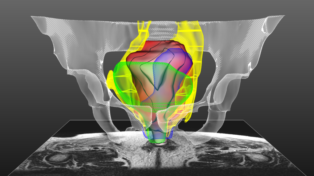
This image depicts PelVis [1], an interactive application for surgical planning for the Total Mesorectal Excision (TME) procedure. During this surgical procedure, undesired side-effects occur in up to 80% of the cases due to damage to the autonomic nerves. These nerves are damaged easily, since they are not visible in pre-operative MRI or even during the surgery. In order to visualize these nerves, we built an atlas model, the Virtual Surgical Pelvis (VSP) [1], that reveals zones in which the autonomic nerves reside based on cryosection and immunohistochemical studies. In the PelVis application, we register this atlas to patient-specific clinical MRI data and thus are able to make patient-specific virtual models of the individual patient, and to reveal the autonomic nerve zones pre-operatively, as displayed here in yellow. We highlight the distance of the mesorectal wall to these nerve zones using a colormap (red to white) combined with isolines. Furthermore, other surgically relevant anatomy is shown for spatial context, without occluding the view on the mesorectum, and the linked atlas-enriched MRI data can be explored interactively [3].
[1]: Smit, N., Lawonn, K., Kraima, A., DeRuiter, M., Sokooti, H., Bruckner, S., … & Vilanova, A. PelVis: Atlas-based Surgical Planning for Oncological Pelvic Surgery. (2017) IEEE Transactions on Visualization & Computer Graphics, (1), 1-1. Accepted, to appear.
[2]: Kraima, A., Smit, N. N., Jansma, D., West, N. P., Quirke, P., Rutten, H. J., … & DeRuiter, M. C. (2014). 62. The virtual surgical pelvis: A highly-detailed 3D pelvic model for anatomical education and surgical simulation. European Journal of Surgical Oncology, 40(11), S32.
 I know, it’s been a while since I last posted, and it’s also been a while since this happened, but… I was one of the winners of the MedViz image contest 2016 which was given out during the joint MedViz conference and Eurographics Workshop on Visual Computing in Biology and Medicine (aka VCBM), right here in Bergen.
